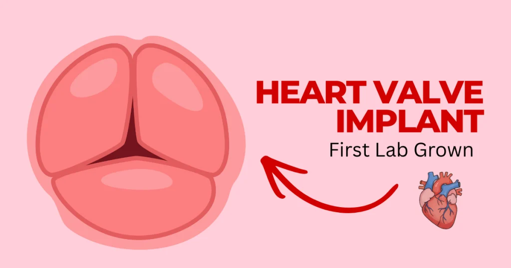
The “Bio-Scaffold” Miracle: First Successful 3D-Bioprinted Heart Valve Implants
The medical world just shifted on its axis. Today, researchers at the Zurich Institute announced a milestone that feels like science fiction but is very much our new reality: the first successful implantation of a 3D-bioprinted heart valve created from a patient’s own cells.
As a healthcare professional, I’ve seen the limitations of our current “gold standards.” Whether we use mechanical valves or tissue valves from pigs or cows, there has always been a “catch.” Mechanical valves require a lifetime of grueling blood thinners; animal valves eventually wear out and fail.
But a living valve? One that heals, grows, and breathes with the patient? That is the “Bio-Scaffold” miracle.
Why Current Solutions Fall Short
To appreciate why this breakthrough matters, we have to look at the patients who suffer most under the current system: children and young adults.
- The Growth Gap: If a five-year-old receives a mechanical or animal valve, that valve stays the same size while the child grows. This leads to a cycle of high-risk open-heart surgeries every few years to “upsize” the hardware.
- The Anticoagulation Burden: Mechanical valves are prone to blood clots. Patients must take Warfarin (blood thinners) daily, which requires constant blood monitoring and restricts physical activity due to the risk of internal bleeding.
- The Degradation Timer: Bioprosthetic (animal) valves usually last only 10 to 15 years in adults—and even less in younger, more active patients.

The Zurich Breakthrough: How It Works
The team at the Zurich Institute didn’t just build a valve; they built a biological blueprint. Here is the step-by-step process of this medical marvel:
- Stem Cell Harvest: Doctors take a small sample of the patient’s own stem cells (usually from bone marrow or fat tissue).
- The Bio-Ink Formulation: These cells are cultured and mixed with a specialized “bio-ink”—a gelatinous material that mimics the body’s natural extracellular matrix.
- 3D Precision Printing: Using a high-resolution bioprinter, the “Bio-Scaffold” is printed layer by layer to match the exact anatomical dimensions of the patient’s heart, captured via 3D imaging.
- The “Living” Integration: Once implanted, the body recognizes the scaffold as “self” rather than a foreign object. The patient’s cells begin to populate the scaffold, eventually replacing the printed material with natural, living heart tissue.
A Permanent Solution for the Next Generation
This technology marks the beginning of the end for “re-do” surgeries. Because the valve is made of living tissue, it possesses remodeling capabilities. It can expand as a child grows. It can repair itself if minor cellular damage occurs.
For a young patient, this means one surgery instead of five. It means a life free from the “click-click” sound of a mechanical valve and the constant worry of blood thinner levels.
“We aren’t just giving them a new part,” says the lead researcher at Zurich. “We are giving them their own tissue back, organized in a way that saves their life.”
The Road Ahead: From Concept to Standard Care
While today’s news is a landmark success, we are at the dawn of this transition. The primary focus now is long-term monitoring to ensure these valves maintain their structural integrity over decades. However, the initial data suggests that the integration of the “Bio-Scaffold” is seamless, with zero signs of rejection—a feat impossible with traditional transplants.
Health Disclaimer
This article is for informational purposes only and does not constitute medical advice, diagnosis, or treatment. Always seek the advice of your physician or other qualified health provider with any questions you may have regarding a medical condition. Never disregard professional medical advice or delay in seeking it because of something you have read on this website. DrugsArea
Sources & References
- Zurich Institute of Regenerative Medicine tag:Zurich-Institute
- World Health Organization: Cardiovascular Diseases tag:WHO-Health
- Journal of Biomedical Materials Research tag:Medical-Research
- The Lancet: Innovations in Cardiac Surgery tag:The-Lancet
People Also Ask
1. What is a “Bio-Scaffold” 3D-printed heart valve?
A bio-scaffold is a temporary, 3D-printed framework made of specialized “bio-ink”—a mix of biocompatible polymers and, in some cases, the patient’s own living cells. Unlike traditional metal or plastic valves, this scaffold acts as a “mold” that encourages your body to regrow its own natural heart tissue. Over time, the scaffold safely dissolves, leaving behind a functional, living valve made entirely of your own cells.
2. Is the 3D-printed heart valve permanent or will it dissolve?
The scaffold itself is designed to be bioresorbable, meaning it eventually dissolves (typically within 6 to 12 months). However, the results are permanent. Because the scaffold triggers your body to build a replacement valve out of native tissue, you aren’t left with a foreign object in your heart; you’re left with a “new” version of your own original anatomy.
3. Do I still need to take blood thinners with a bioprinted valve?
One of the biggest breakthroughs of the bio-scaffold “miracle” is the potential to eliminate lifelong blood thinners. Traditional mechanical valves require anticoagulants (like Warfarin) to prevent clots on the metal surfaces. Because bioprinted valves eventually become your own living tissue, the body recognizes them as “self,” significantly reducing the risk of rejection and blood clots.
4. Why is this technology called a “miracle” for pediatric patients?
For children born with heart defects, traditional valves are a nightmare because they don’t grow. This leads to multiple high-risk open-heart surgeries as the child gets older. A 3D-bioprinted valve grows with the patient. Since it’s made of living cells that divide and expand, a single implant can potentially last a lifetime, sparing children from a childhood of repeated operations.
5. How long does the 3D-bioprinting process take for a custom valve?
While the research took decades, the actual printing is remarkably fast. Using advanced Digital Light Processing (DLP), a custom-fitted heart valve can be printed in as little as 120 seconds. However, the “pre-op” phase—which involves taking a CT scan of your heart and processing your stem cells into bio-ink—typically takes a few weeks.
6. Can my body reject a 3D-bioprinted heart valve?
The risk of rejection is significantly lower than with pig (porcine) or cow (bovine) valves. By using autologous cells (cells harvested from your own skin or blood), the bioprinter creates a valve that matches your unique genetic blueprint. Your immune system views the implant as part of your body rather than a foreign invader.
7. How is a bio-scaffold valve implanted—is it open-heart surgery?
Not necessarily. Many of these new scaffolds are made from shape-memory polymers. These allow the valve to be “folded” into a thin tube and delivered via a minimally invasive catheter (similar to a TAVR procedure). Once it reaches the heart and hits body temperature, it “remembers” its shape and unfolds into place, avoiding the need to crack the chest open.
8. How does a bioprinted valve compare to a pig or cow valve?
Biological valves from animals (xenografts) usually wear out and calcify after 10–15 years, requiring a second replacement. 3D-bioprinted valves are designed to self-repair and remodel, just like a healthy human valve. This “living” quality means they are theoretically much more durable and less likely to stiffen or fail over time.
9. Is this technology available at all hospitals yet?
As of 2026, we are in the “early adopter” phase. While the first successful human implants have made headlines, the procedure is currently limited to leading cardiac research centers and patients enrolled in specific clinical trials (like the FDA Breakthrough Devices Program). It is expected to become more widely available over the next 5 to 10 years.
10. What are the risks of 3D-bioprinted heart valves?
As with any new “miracle” tech, the primary risk is long-term data. While early trials are incredibly promising, doctors are still monitoring how these “regrown” valves hold up after 20 or 30 years of use. There are also technical risks, such as the scaffold dissolving too quickly before the new tissue is strong enough to handle the heart’s high pressure.